Some color changes in reptiles
are morphological rather than a physiological color change. Some reptiles are
able to experience color change but it is not instantaneous. Conversely other
reptiles can vigorously change color using specialized signaling mechanisms in
order to change color quickly which is also dependent on the biotic or abiotic
indication (Stuart-Fox &Adnan Moussalli, 2008).
What Do Reptiles Use to Camouflage?
Reptiles mainly use
pigmented containing cells called chromatophores in a specific orientation for
color change which is sometimes referred to as the dermal chromatophore
component. There are different kinds of chromatophores depending on the
species.
Where Are These Cells Located And What Are The Common Types?
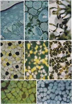 Figure shows from left to right: skin adapted to white background, melanosomes surrounding iridophores, yellow spots over iridophores, same as before except activated melanosomes, melanophores that are widespread, adapted to light background -- green combined effect of xanthophore and iridophores, and skin with xanthophores removed leaving iridophores.
The chromatophores
cells are located in the dermis. The most common cells found in reptiles
include the Xanthophores which are yellow, the Melanophores which are black and
or brown, and the Erythophores which are red.
For example, if a reptile is prompted to change to a darker color the melanophores will move to cover other chromatophores and absorb light from iridopohores. The melanophores are found underneath or between other colored cells, such as xanthophores, which are found above iridophores (Kuriyama et. al. 2006). Melanophores are generally more active than the other chromatophores. While color-producing cells are stationary, melanophores, once stimulated, will secrete melanosomes, which move to "cover" the color-producing cells above. A gradient of melansomes occurs, showing different intensities of color. The secretion of melanosomes also causes the melanophore to expand, blocking more light (Goldman 1969; Bagnara et al. 1968).
For example, if a reptile is prompted to change to a darker color the melanophores will move to cover other chromatophores and absorb light from iridopohores. The melanophores are found underneath or between other colored cells, such as xanthophores, which are found above iridophores (Kuriyama et. al. 2006). Melanophores are generally more active than the other chromatophores. While color-producing cells are stationary, melanophores, once stimulated, will secrete melanosomes, which move to "cover" the color-producing cells above. A gradient of melansomes occurs, showing different intensities of color. The secretion of melanosomes also causes the melanophore to expand, blocking more light (Goldman 1969; Bagnara et al. 1968).
The Iridophores
Iridophores are found in the next layer under
the chromatophores (Hanlon et. al. 1990; Cooper et. al. 1990). Iridophores are
also used to reflect light into the colored cells. The light enters the skin
and is reflect when it makes contact with iridophores. Iridophores work by
reflecting light and can also be used to mask organs, as is often the case with
the silver coloration around the eyes and ink sacs. In addition they can assist
in concealment and communication. Iridophores are layered stacks of platelets
that are chitinous in some species and protein based in others. They are responsible
for producing the metallic looking greens, blues and golds seen in some
species, as well as the silver color around the eyes and ink sac of others
(Hanlon & Messenger, 1996). As a result, there is usually a particular
structure used to produce a color.
Physiological Pathways of Camouflage in Reptiles
The two main
physiological pathways include the hormonal or the neural response to
stimulation. However there are some species that use a combination of both
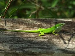
pathways. For example, in the species of
lizard Anolis carolinensis, color change or camouflage occurs when a stimulated
pituitary gland secrets melanoctyte stimulating hormone (MSH) which will then
activate the melanophore. In other species for instance the chameleons,
melanophores are directly stimulated by the nervous system which usually
incorporats the neurotransmitter, norepinephrine (Taylor & Hadley, 1970;
Goldman 1969). The fact that neural response is faster than hormonal means that
color change will occur faster in reptiles that use such a pathway (Stuart-Fox
et. al. 2008). The release of MSH or neurotransmitters is the result of many
factors which can includes stress, illumination, and temperature. In the case
of illumination, some reptiles such as anoles detect light via their eyes but
also with photoreceptors on their skin. Generally speaking, the warmer it is,
the lighter the reptile will be in order to absorb less light and therefore
heat (Taylor & Hadley, 1970; Macedonia, 2001).
